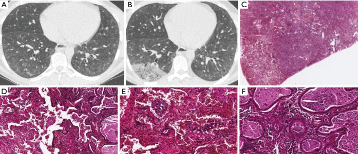
Figure S1.
(A) CT scan of a 34-year-old man with ILD in a context of scleroderma in 2015; (B) CT scan of the same patient in 2017 showing a new right lower lobe opacity; (C) presence of a tumoural nodule of adenocarcinoma in a case of pulmonary involvement by scleroderma. ×1, HES; (D) adenocarcinoma is present in the right of the field and non tumoural lung parenchyma on the left, with NSIP. ×10, HES; (E) lung parenchyma with NSIP. The witness of the parietal wall is increased and there is inflammatory cells and alveolar hemorrhage. ×20, HES; (F) invasive acinous adenocarcinoma with inflammatory cells. ×20, HES. NSIP, non-specific interstitial pneumonia; ILD, interstitial lung disease; CT, computed tomography.

