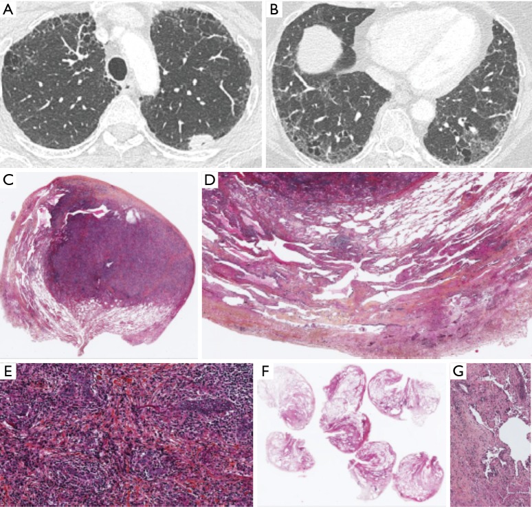
Figure S2.
(A) CT scan of a 52-year-old tobacco smoker woman with a left upper lobe nodule. Presence of subpleural reticulation and paraseptal emphysema; (B) CT scan of the lower lobes showing a predominance of peripheral and basal predominance of reticular abnormalities with emphysema. The radiological pattern is possible UIP; (C) lymphoepithelial carcinoma in a case of UIP. ×1, HES; (D) lung parenchyma surrounding the nodule with peripheral subpleural heterogeneous fibrosis. ×10, HES; (E) lymphoepithelial carcinoma with carcinomatous islands associated with fibrosis and a lot of inflammatory cells. ×20, HES; (F) heterogeneous fibrosis with subpleural and peripheral predominance; (G) fibroblastic focus. CT, computed tomography; UIP, usual interstitial pneumonia.

