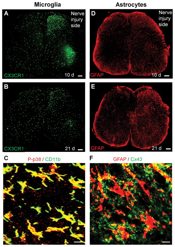
Figure 3.
Distinct and time-dependent activation of microglia and astrocytes in the spinal cord after nerve injury. (A, B) Microglia activation revealed by increased CX3CR1 expression in the spinal cord 10 days (A) and 21 days (B) after nerve injury in Cx3cr1-GFP mice. Scale, 100 μm. (C) Phosphorylation of p38 MAP kinase (P-p38) in CD11b+ microglia in the spinal cord dorsal horn 7 days after nerve injury. Scale, 20 μm. (D, E) Astroctye activation revealed by increased GFAP expression in the spinal cord 10 days (D) and 21 days (E) after nerve injury in mice. Scale, 100 μm. (F) Expression of Cx43 in GFAP+ astrocytes in the spinal cord dorsal horn 21 days after nerve injury. Scale, 20 μm. All the images have not been published.

