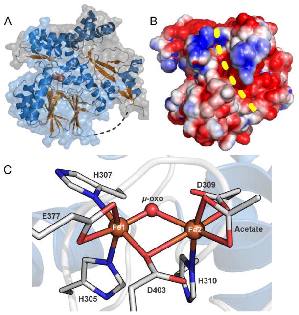
Figure 2.
X-ray crystal structure of CmlA (A) Structure of diferric CmlA at 2.17 Å resolution. The N- and C-terminal domains are shown in gray and light blue respectively. (B) Space-filling surface representation showing the surface groove (yellow dashed line) formed at the interface of the N- and C-terminal domains. The active site cavity extends into the protein from the top end of the groove as shown. (C) Ligand environment of the diferric diiron cluster, PDB 4JO0. In the X-ray crystal structure of the diferrous enzyme, PDB 5KIK, the acetate ligand on Fe2 is replaced by two waters. Adapted with permission from T. M. Makris, C. J. Knoot, C. M. Wilmot and J. D. Lipscomb, Biochemistry, 2013, 52, 6662–6671. Copyright 2013 American Chemical Society and A. J. Jasniewski, C. J. Knoot, J. D. Lipscomb and L. Que, Jr., Biochemistry, 2016, 55, 5818–5831. Copyright 2016 American Chemical Society

