
Figure 1. Ketamine increases cell proliferation in the hippocampal granule cell layer and subgranular zone after CCI.
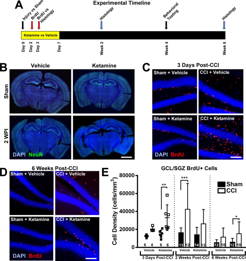
(A) Experimental timeline. Adult mice were randomized to undergo controlled cortical impact (CCI) or Sham injury, followed by continuous systemic administration of either Ketamine or Vehicle for one week, beginning on Day 0. Mice received the mitotic marker BrdU on days 2 and 3 after injury to permanently label actively dividing cells. Immunohistochemical analysis of the ipsilateral hippocampus was performed in separate cohorts of mice at three different time points: 3 days, 2 weeks and 6 weeks post-intervention, to distinguish patterns in cell proliferation and differentiation early and delayed stages. Mice in the 6-week arm underwent behavioral testing in weeks 4 and 5 post-intervention. (B) Representative images of coronal sections from Sham (non-injured) and CCI-injured mice at 2 weeks post-injury (WPI). Scale bar = 2mm. (C) Representative higher power images of the ipsilateral hippocampal dentate gyrus from injured and Sham-treated mice that received ketamine or vehicle after CCI. Ketamine administration after CCI induced a robust increase in newly born (BrdU+, red) cells in the granule cell layer and subgranular zone (GCL/SGZ, visualized with DAPI, blue) 3 days after injury. Scale bar = 150 μm. (D) Images of BrdU-stained dentate gyrus taken 6 weeks after CCI, with BrdU administered 2-3 days after CCI in the presence of ketamine or vehicle treatment. Scale bar = 150 μm. (E) Two-way ANOVA analysis of BrdU+ labeled cell density in the GCL/SGZ at the various time points evaluated in this study. Bars represent mean cell densities (cells/mm3) and error bars represent the standard deviation (SD); numbers inside bars represent the N for each group. For the three-day timepoint, individual values are displayed as well as the mean/SD. *p < 0.05; **p < 0.01; ***p < 0.001.