
Figure 5. Ketamine Treatment Facilitates Rapid Microgliogenesis after CCI.
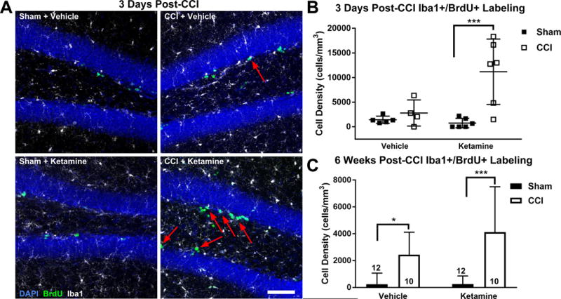
(A) Representative images of the GCL (DAPI, blue), microglia (Iba1+, white) and newly generated BrdU-labeled cells (green) 3 days after CCI or sham treatment. Newly generated microglia (Iba1+/BrdU+) are designated by arrows. Scale bar = 150 μm. (B) Quantification of microgliogenesis 3 days after injury demonstrated a significant increase in newly born microglia within the GCL/SGZ only in the Ketamine + CCI group. (C) At 6 weeks post-intervention, microglia born after CCI/Sham were significantly increased in both CCI groups versus their respective Sham groups. Bars represent mean cell densities (cells/mm3) and error bars represent SD; numbers inside bars represent the N for each group; * p < 0.05; ***p < 0.001.