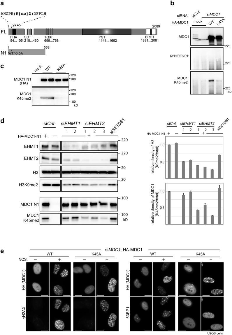
Figure 3.
EHMT2 promote methylation of MDC1 at lysine 45. (a) Schematic illustration of the full-length (FL) and deletion mutant (N1) of MDC1. Asterisk indicates the position of lysine 45 (wild type; WT or the alanine-substituted mutant; K45A). FHA, forkhead-associated domain; SDT, Ser-Asp-Thr repeats; TQXF, Thr-Gln-X-Phe repeats; PST, Pro-Ser-Thr repeats; BRCT, tandem BRCT domain. (b–d) Immunoblotting analysis of whole-cell extracts from U2OS cells transfected with (b) HA-tagged MDC1 (WT or K45A) and control or MDC1-targeting siRNAs, (c) HA-tagged MDC1-N1 (WT or K45A), and (d) HA-tagged MDC1-N1 (WT) and control, two EHMT1-targeting siRNAs (EHMT1 siRNA-1 and siRNA-2) or three EHMT2-targeting siRNAs (EHMT1 siRNA-1 siRNA-2 and siRNA-3; see methods), using the indicated antibodies. Quantification of methylation levels in histone H3 and MDC1 shown in right panels. Relative density was calculated based on signal intensities normalized against levels of total histone H3 (upper panel) and total MDC1-N1 (lower panel). Error bars, SD from triplicate measurements. (e) Immunofluorescence analysis of U2OS cells transfected with HA-tagged MDC1 (WT or K45A) and MDC1-targeting siRNA, and co-immunostained with indicated antibodies at 2 h after exposure to neocarzinostatin (NCS, 50 ng/ml for 15 min). A representative image of each treated or control cells is shown, as indicated. Scale bar, 10 μm.

