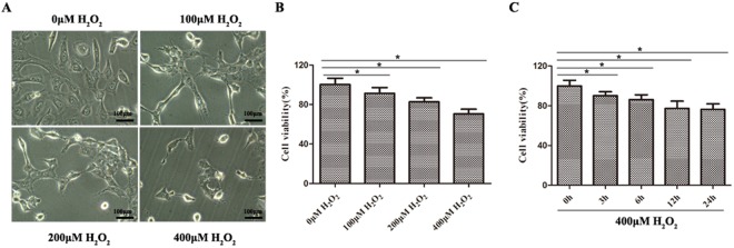
Figure 1.
Oxidative damage model induced by H2O2was established in GES-1 cells. (A) Morphological changes in GES-1 cells exposed to 4 different concentrations of H2O2, including 0 μM, 100 μM, 200 μM and 400 μM. (B) The effects of various H2O2 concentrations on cell viability in GES-1 cells, as determined by MTT assay. The cell viability was gradually decreased in a dose-dependent manner; *P < 0.05. (C) The effects of 400 μM H2O2 on cell viability in GES-1 cells for 0 h, 3 h, 6 h, 12 h and 24 h. The cell viability gradually declined in a time-dependent manner; *P < 0.05.

