Figure 1.

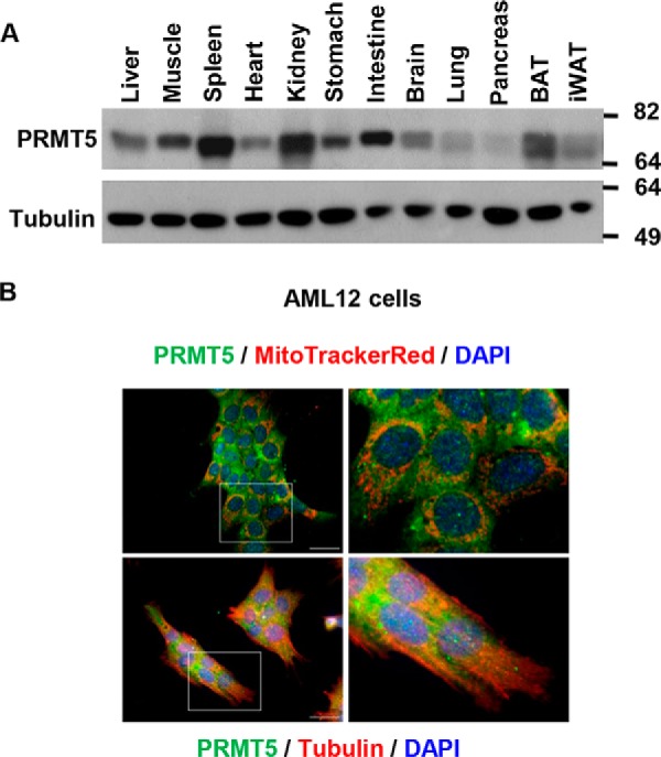
PRMT5 is ubiquitously expressed in mouse tissues. A, PRMT5 protein levels in mouse tissues were determined by Western blot analysis. BAT, brown adipose tissue; iWAT, inguinal WAT. B, localization of PRMT5 in the mouse AML12 liver cell line. PRMT5 expression was visualized by immunofluorescent staining (green), the cytoskeleton was labeled by immunofluorescent staining of tubulin (red), and nuclei were stained by DAPI (blue). Scale bars = 200 μm. All images represent three independent experiments.
