
Figure 4.
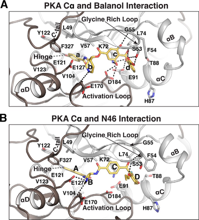
Structural comparison between PKA Cα bound with balanol and N46. A and B, detailed interactions between PKA Cα and balanol (PDB code 1BX6) (A) orN46 (B). Only the regions near the active site are shown. Residues contacting balanol or N46 are shown as sticks. Direct hydrogen bonds are shown as dotted lines. The rings of balanol and N46 are labeled a–d and A–D, respectively.