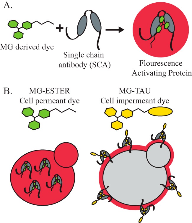
Figure 2.

The use of a fluorogen-activating protein to report on cellular residence. A, the FAP technique makes use of a MG-derived dye that binds an SCA. Neither the MG nor the SCA is fluorescent, but when the MG dye is bound by an SCA, fluorescence is detected. B, the MG-derived dye can be conjugated to a membrane-soluble side chain (MG-ESTER, cell-permeant dye) that freely passes through the yeast cell wall and the plasma membrane, where it can be bound by intracellular SCAs to activate fluorescence and monitor the intracellular levels of a tagged protein. The MG-derived dye can also be conjugated to a membrane-impermeant side chain, as is the case with the MG-B-TAU dye. This dye can no longer enter the cell, so only the SCAs on the external face of the cell surface will be bound by dye and fluoresce.
