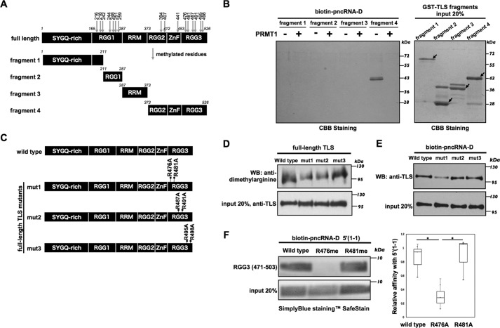
Figure 2.
The Arg-476 residue contributes to the pncRNA-D–binding site. A, TLS methylation sites and the fragments. TLS contains a total of 16 arginine residues (arrows) that can be methylated. B, biotinylated pncRNA-D was pulled down with unmethylated or methylated TLS fragments 1–4. The pncRNA-D–bound TLS fragments were detected by Coomassie Brilliant Blue staining; 20% of the protein was loaded as input. C, GST-TLS constructs with the substitution of arginine by alanine are indicated as mut1 (R476A and R481A), mut2 (R487A and R491A), and mut3 (R495A and R498A). D, arginine methylation levels of GST-TLS mutants (mut1, mut2, and mut3) were detected by an anti-dimethylarginine antibody. E, biotinylated pncRNA-D was incubated with TLS mutants (mut1, mut2, and mut3). The pncRNA-D–bound TLS was detected by an anti-TLS antibody. F, biotinylated pncRNA-D fragment 5′(1-1) was incubated with synthesized peptides (RGG3, TLS amino acids 471–503) with one of their arginine residues methylated (R476me and R481me). Samples were stained with the SimplyBlueTM SafeStain for analysis. The intensity of the bands was measured by ImageJ and applied in the box plot analysis. n = 3. *, p < 0.05. 20% of the protein used for RNA pulldown assays was loaded as input.

