Figure 6.
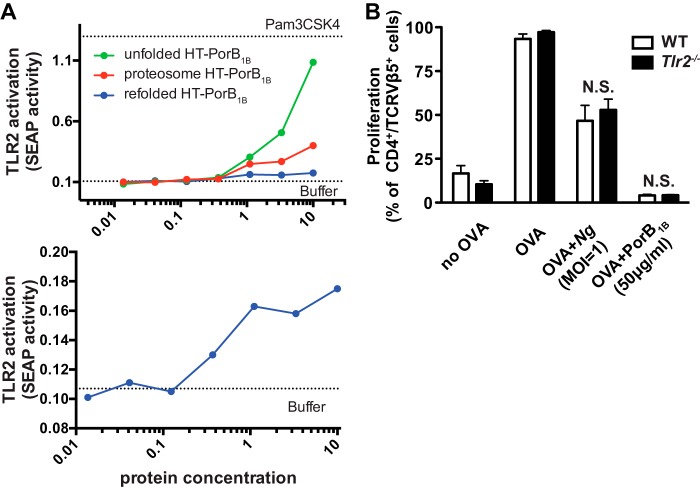
PorB1B does not inhibit dendritic cell–induced T cell proliferation through TLR2. A, HEK-Blue hTLR2 reporter cells were treated with LDAO-containing buffer (0.0075%) alone or with the indicated concentrations of recombinant unfolded, refolded, and proteosome HT-PorB1B. After 24 h, cell culture supernatants from the treated cells were assayed for the activity of secreted alkaline phosphatase as described under “Experimental procedures.” The top panel shows the dose response for all three PorB1B preparations, whereas the bottom panel shows only refolded PorB1B with an expanded y axis. B, WT or Tlr2−/− dendritic cells were treated with GWM (no OVA), OVA, OVA + Ng (m.o.i. = 1), or OVA + PorB1B (50 μg/ml) and then co-cultured with CSFE-labeled OT-II T cells. T cell proliferation was assessed by flow cytometry. The mean ± S.E. (n = 4) of the percentage of OT-II T cells that underwent proliferation over 7 days is plotted for cells co-cultured with WT dendritic cells (black columns) or Tlr2−/− dendritic cells (white columns) for the indicated treatments. Statistical significance was assessed by two-way ANOVA with a post hoc Bonferroni analysis for multiple comparisons. No statistically significant difference was found between WT and Tlr2−/− cells.

