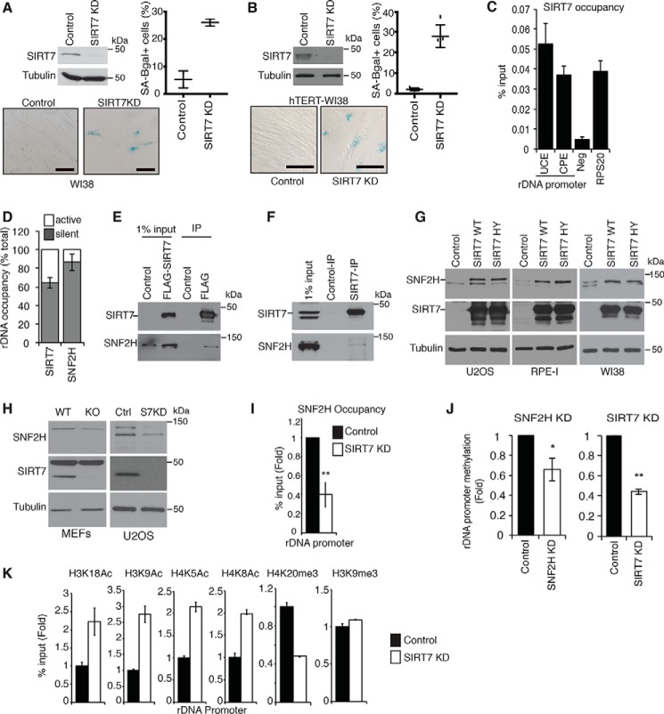
Figure 1.
SIRT7 maintains rDNA heterochromatin and guards against cellular senescence. A and B, SA-β-gal assays showing increased senescence following SIRT7 KD in WI38 (A) or hTERT-immortalized WI38 (B) cells. Scale bars, 100 μm. Data are average of two to five fields ±S.D. (error bars). Immunoblots of SIRT7 protein and representative SA-β-gal images are shown. C, ChIP-qPCR showing SIRT7 occupancy at rDNA promoter regions upstream promoter element (UCE) and core promoter element (CPE). Neg, intergenic negative control; RPS20, positive control. D, ChIP-CHOP analysis showing percentage of rDNA-bound SNF2H or SIRT7 present at silent versus active rDNA copies. E and F, immunoblots showing coimmunoprecipitation of SNF2H with FLAG-SIRT7 expressed in U2OS cells (E) or endogenous SIRT7 IPs from 293T cells (F). Anti-FLAG antibodies were used for negative control IPs. G, immunoblots showing increased SNF2H levels in the indicated cell lines overexpressing WT or catalytically inactive (H187Y) SIRT7 proteins. H, immunoblots showing decreased SNF2H protein levels in SIRT7 KO MEFs and SIRT7 KD U2OS cells. I, ChIP-qPCR showing decreased SNF2H occupancy at rDNA promoters in SIRT7 KD U2OS cells. J, decreased DNA methylation at rDNA promoters in SIRT7 KD or SNF2H KD cells. K, ChIP-qPCR showing changes in histone acetylation and methylation marks at rDNA promoters in SIRT7 KD cells. In I and J, * indicates p < 0.05, and ** indicates, p < 0.01 (one-tailed Student's t test). In all graphs, data show averages of three technical replicates ±S.E. (error bars) and are representative of two to four biological replicates.

