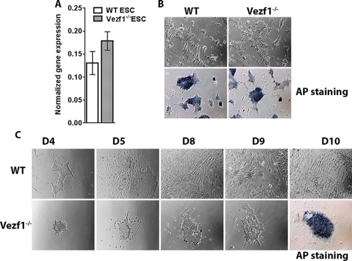
Figure 2.
A, gene expression analysis by RT-qPCR of Oct4 in WT and Vezf1−/− ESCs. B, alkaline phosphatase (AP) staining for pluripotency in WT and Vezf1−/− ESCs; the presence of dark blue stain indicates positive for pluripotency. C, WT and Vezf1−/− ESCs were differentiated using 10 ng/μl of VEGF-A165 for 5, 6, 7, and 10 days and visualized using brightfield microscopy at ×10 magnification. Unlike the WT cells, the Vezf1−/− cells were unable to differentiate and most of the cells died. The field view is the representation of proliferation during differentiation and cell number. The panel for D10 shows alkaline phosphatase staining. A strong signal of Vezf1−/− cells indicates presence of undifferentiated stem cells. WT, wildtype ESCs; Vezf1−/−, Vezf1 knockout ESCs; UD, undifferentiated; D4–D10, days post-differentiation.

