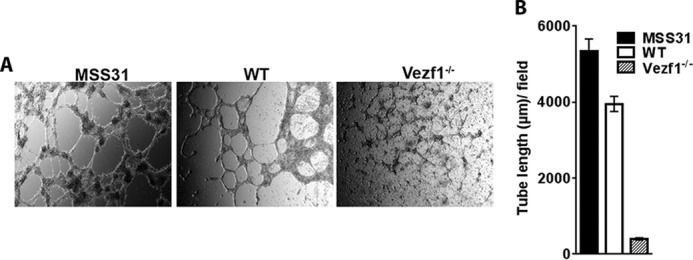
Figure 5.

A, differentiated WT and Vezf1−/− EC plated in VEGF-supplemented Matrigel were incubated at 37 °C for 5–15 h. The formation of tube structures is visualized by brightfield microscopy. Mouse endothelial cell line MSS31 is used as a positive control. The images were taken at ×10 magnification at 12 h for MSS31, and 6 h for WT and Vezf1−/−. B, measurement of tube length using ImageJ software. Compared with MSS31 and WT ECs, Vezf1−/− ECs were unable to make tube-like structures in Matrigel. WT, wildtype; Vezf1−/−, Vezf1 knockout.
