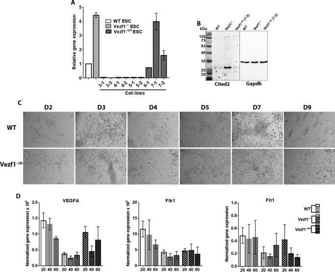
Figure 6.
A, gene expression analysis of Cited2 by RT-qPCR in Vezf1−/sh cell lines. Change in gene expression was plotted relative to that of WT ESCs, set to 1. Labels on the x axis represent various stable cell lines, of which the Vezf1−/sh cell line (7-2) has Cited2 expression reduced to the levels similar to WT ESCs. B, Western blot analysis using 50 μg of total cell extract from the WT, Vezf1−/−, and Vezf1−/sh (7-2) ESCs probed with anti-Cited2 antibody and anti-GAPDH. C, differentiation of WT, Vezf1−/−, and Vezf1−/sh (7-2) ESCs was induced using 20 ng/μl of VEGF-A165. D2–D6 are days post-differentiation. Compared with the differentiating WT cells, Vezf1−/sh (7-2) show similar morphology and cell number indicating at least a partial rescue of their ability to differentiate into ECs. D, gene expression analysis of Vezf1−/sh (7-2) ESCs, which were differentiated to ECs at 20, 40, and 60 ng/μl of VEGF-A165. Expression of pro-angiogeneic genes VEGF-A, Flk1, and Flt1 was measured. Compared with WT and Vezf1−/− cells, gene expression was largely rescued in Vezf1−/sh (7-2) ECs. The data represent the average and S.D. of 3 to 4 replicates. WT, wildtype ESCs; Vezf1−/−, Vezf1 knockout ESCs; Vezf1−/sh, stable transgenic Vezf1−/− ESCs expressing Cited2-shRNA; 20, 40, 60, ng/μl of VEGF-A165 used for differentiation.

