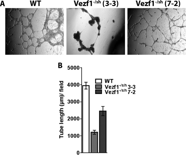
Figure 7.

A, WT, Vezf1−/sh (7-2), and Vezf1−/sh (3-3) cells were differentiated for 10 days and used in a tube-formation assay. The Vezf1−/sh cell line (3-3) had about 7-fold lower Cited2 expression than WT. Compared with the WT cells, tube formation was rescued in Vezf1−/sh (7-2) ECs, which was absent in Vezf1−/sh (3-3) ECs. Images were taken at 12 h for Vezf1−/sh (3-3) and 6 h for WT and Vezf1−/sh (7-2). B, tube length was measured by ImageJ software and plotted.
