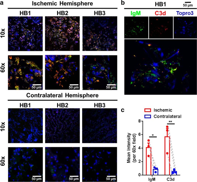
Figure 10.
Complement and IgM deposition in the ischemic hemisphere of human patients who died from acute stroke. a, Immunofluorescence microscopy showing IgM (green) and C3d (red) deposition in the ischemic but not contralateral hemisphere of patients who died from acute stroke. HB1, 24 h poststroke; HB2, 24–48 h poststroke; HB3, 48 h poststroke; blue, DAPI. b, High-resolution 3D rendering of IgM and C3d deposition showing colocalization of C3d and IgM in the ischemic penumbra. c, Pairwise comparisons of C3d and IgM deposition in the ipsilateral compared with the contralateral hemisphere showing significantly higher levels in the ipsilateral hemisphere of acute stroke patients. Pairwise two-way ANOVA, Bonferroni's correction, n = 3, *p < 0.05, **p < 0.01.

