Figure 8.
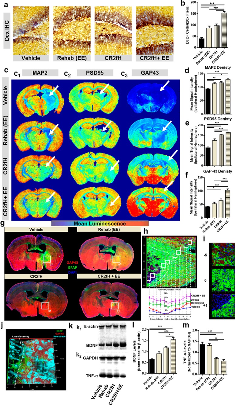
CR2fH removes the inhibition on regenerative mechanisms promoting the outcome of rehabilitation therapy. a, Dcx immunostaining of perilesional hippocampi quantified in b showing a significant increase in the number of neuroblasts migrating to the ipsilateral hippocampus 15 d after injury with rehabilitation, CR2fH treatment, or combination therapy. However, combination of CR2fH and rehabilitation showed a more robust increase in Dcx+ cells compared with either single intervention. ANOVA. n = 6 animals (2 fields each), ***p < 0.001. c–f, Immunostaining for markers of regeneration and remodeling, including dendritic arborization (MAP2), synaptic density (PSD-95), and axonal growth (GAP-43) of full-brain slices showing that a combination of CR2fH and rehabilitation resulted in the most pronounced and significant increase in dendritic and axonal growth to the perilesional area with subsequent increase in synaptic density. Significant increase in regenerative markers was also seen in CR2fH-treated animals (all three markers) and in rehabilitation-only animals (MAP2 and PSD95). ANOVA, full hemispheres quantified, *p < 0.05, **p < 0.01, ***p < 0.001. Heat map shows mean luminescence. g, Costaining for GAP-43 and GFAP showing that robust astrogliosis inhibits the regrowth of regenerating (GAP-43+) axons into the perilesional area. CR2fH significantly reduced astrogliotic scarring, resulting in increased GAP-43 infiltration to perilesional brain, whereas CR2fH in combination with rehabilitation further increased the levels of GAP-43 in the perilesional areas. Rehabilitation-only animals showed limited GAP43 increase that was still blocked by astrogliosis. h, Quantification of GAP-43 density in the perilesional area showing that vehicle and rehabilitation animals showed a significant reduction in GAP43 density surrounding the areas of astrogliosis compared with CR2fH with or without rehabilitation. p < 0.01 comparing vehicle or rehabilitation with CR2fH with or without rehabilitation at locations −2 to 3. Two-way ANOVA, n = 3/group. i, Representative 3D-rendered fields from h at different positions relative to the astrogliotic scar. j, Notable reduction in GAP43 was observed near the areas of astrogliosis in vehicle-treated animals despite comparable neuronal density across the boundaries of the scar (NeuroTrace, cyan). k, Representative Western blots showing levels of neuronal growth factor (BDNF) and TNF-α across the different groups. l, Both CR2fH and rehabilitation alone resulted in a significant increase in the levels of BDNF in the ipsilateral hemisphere compared with vehicle. However, combination therapy resulted in a more robust and significant increase compared with either single intervention. ANOVA, n = 6/group, *p < 0.05, ***p < 0.001. m, Rehabilitation did not influence the levels of TNF-α. However, CR2fH with or without rehabilitation significantly reduced the levels of TNF-α compared with rehabilitation or vehicle groups. ANOVA, n = 5/group, **p < 0.01, ***p < 0.001.

