
Figure 4.
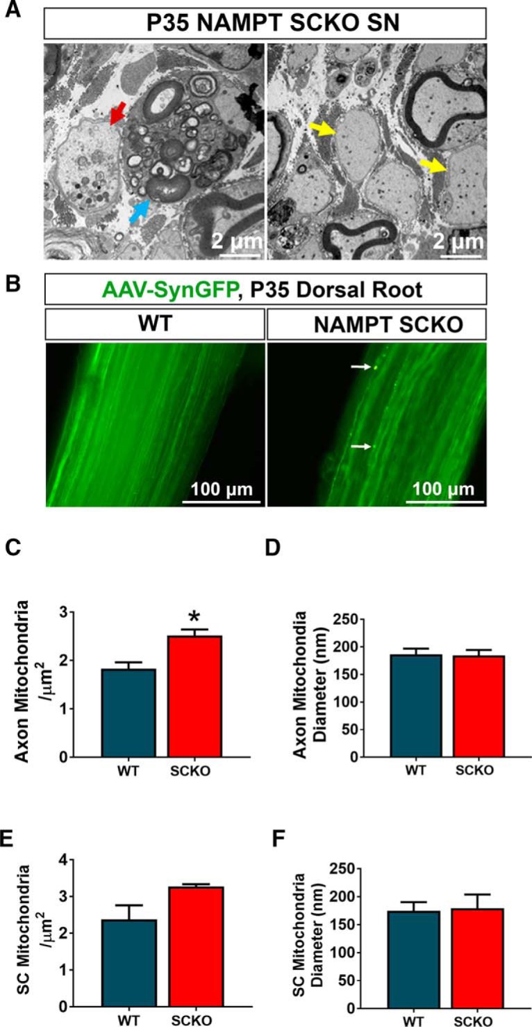
NAMPT SCKO does not induce severe axon degeneration. A, Most axons in NAMPT SCKO mice do not show obvious signs of axon degeneration, but occasional fragmented axons were observed (red arrow). Naked axons (yellow arrows) and myelin-laden macrophages (blue arrow) were frequently observed in NAMPT SCKO nerves. B, DRG neurons were infected with an AAV virus-expressing EGFP driven by the Synapsin promoter. Images shown are whole-mounted dorsal roots. While almost all axons were continuous, dilated axons were occasionally observed in NAMPT SCKO dorsal roots (white arrow). Mitochondria density (C, D) and diameter (E, F) were measured for both axons (C, D) and SCs (E, F) in P35 WT and NAMPT SCKO sciatic nerves using electron microscopic images. *p = 0.02 (unpaired two-tailed Student's t test). n = 3 mice for all experiments in this figure. Data are mean ± SEM.