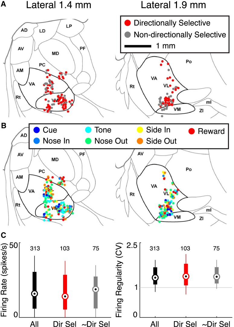
Figure 6.
Anatomical and electrophysiological characteristics of Mthal units. A, Location of directionally (red) and nondirectionally selective (gray) units superimposed on sagittal rat brain atlas images (Paxinos and Watson, 2007). Mthal nuclei [ventral anterior (VA), ventral lateral (VL), ventromedial (VM)] are enclosed within bold lines. AD, Anterodorsal thalamus; AM, anteromedial thalamus; AV, anteroventral thalamus; LD, laterodorsal thalamus; LP, lateral posterior thalamus; MD, mediodorsal thalamus; ml, medial lemniscus; PC, paracentral thalamus; PF, parafascicular thalamus; Po, posterior thalamic nuclear group; Rt, reticular thalamus; ZI, zona incerta. B, Anatomical characterization of Mthal units based on event responsiveness. There was no clear anatomic segregation of units based on directional selectivity or event-responsiveness. C, Left, Median firing rates of all, directionally selective (Dir Sel), and nondirectionally selective (∼Dir Sel) units. Right, Median coefficient of variation (CV) for the same units. Thick lines indicate the 10th to 90th percentiles. The whiskers extend to the most extreme data points not considered outliers.

