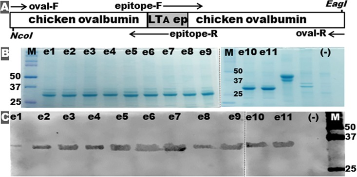
FIG 2.

Construction and detection of epitope-ovalbumin fusion proteins. (A) Illustration of the genetic structure of an epitope-ovalbumin fusion gene, with PCR primers used to insert each LTA epitope into the intron-truncating chicken ovalbumin gene. (B) SDS-PAGE Coomassie blue staining shows each extracted epitope-ovalbumin fusion protein (e1 to e11). (C) Western blot with anti-LT antiserum to characterize each epitope-ovalbumin fusion protein (e1 to e11). Note that e1 to e11 represent epitope-ovalbumin fusion proteins carrying the LTA subunit epitope e1, e2, e3, e4, e5, e6, e7, e8, e9, e10, or e11. “(−)” refers to proteins of host strain E. coli BL21-CodonPlus (DE3). M, molecular marker (Bio-Rad Precision Plus Protein Dual Color Standards), with the 25-, 37-, and 50-kDa bands marked.
