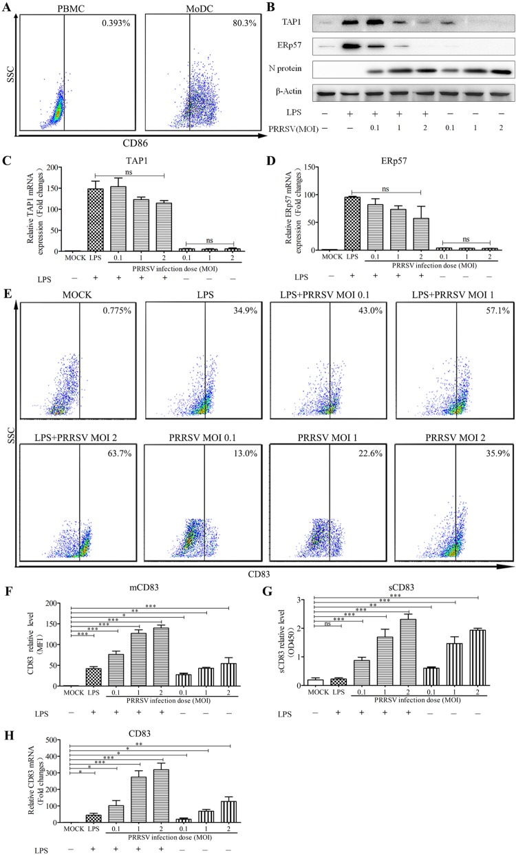
FIG 1.
PRRSV downregulates TAP1 and ERp57 and upregulates CD83 in MoDCs. (A) Porcine monocytes (PMBCs) were cultured for 0 days (left) and 7 days (right) in the presence of GM-CSF and IL-4. After staining with an isotype-matched control antibody, the cells were analyzed for CD86 expression at the cell surface using FACS. (B) MoDCs were infected with PRRSV at an MOI of 0.1, 1, and 2 in the presence or absence of LPS (10 μg/ml) for 24 h. Cell lysates were analyzed for TAP1, ERp57, and N proteins using Western blotting. β-Actin served as a loading control. TAP1 (C) and ERp57 (D) mRNA levels were analyzed by qRT-PCR. mRNA levels were calculated relative to known amounts of template and normalized to β-actin expression. (E) The cells were also analyzed for surface CD83 (mCD83) expression by flow cytometric analysis. (F) Mean fluorescence intensity (MFI) was quantified as a measure of mCD83 production for each analyzed sample. Culture supernatants were also collected, and sCD83 expression was analyzed by ELISA (G) and qRT-PCR (H). MoDCs were inoculated with PRRSV (MOI of 1) at 6, 12, 18, 24, 30, 36, and 48 hpi. All assays were repeated at least three times, with each experiment performed in triplicate. Bars represent means ± SEM from three independent experiments. ***, P < 0.001; **, P < 0.01; *, P < 0.05.

