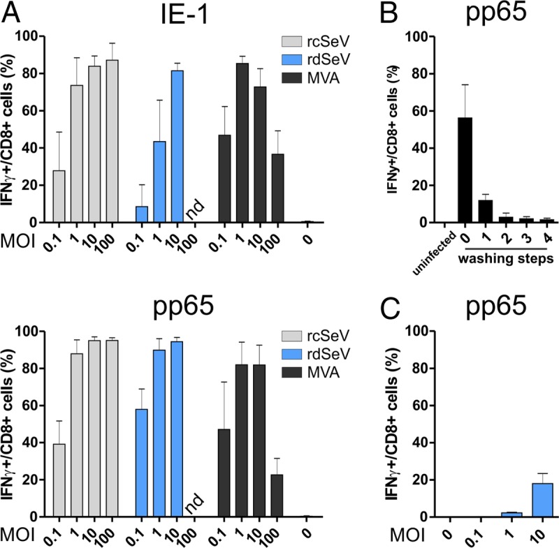
FIG 3.

Infection with both Sendai vectors leads to efficient restimulation of T cells by infected moDCs. (A) Direct presentation assay. moDCs from 3 different HCMV seronegative blood donors were infected at the indicated MOIs with IE-1- or pp65-expressing vectors. At 24 hpi, antigen-specific T cell clones were added at an effector/target ratio of 1:1. After 6 h of cocultivation in the presence of brefeldin A (BFA), cells were stained for CD8 and intracellular IFN-γ and analyzed via flow cytometry. (B) Removal of extracellular virions before cross presentation: HeLa cells were transduced at an MOI of 10 with rdSeV-pp65. At 24 hpi, the supernatant from the overnight culture (lane 0) as well as 4 subsequent washing steps with cell culture medium (lanes 1 to 4) was collected and added to moDCs. Twenty-four h later, a pp65-specific, HLA-matched T cell clone was added to moDCs at an effector/target ratio of 1:1. After 6 h in the presence of BFA, CD8/IFN-γ staining and flow cytometry analysis were performed. (C) Cross-presentation assay. HeLa cells were transduced at the indicated MOIs with rdSeV-pp65. At 24 hpi, cells were washed 4 times and added to moDCs from 3 individual donors at a 1:1 ratio. After 24 h of cocultivation, an antigen-specific T cell clone was added for a HeLa/DC/T cell ratio of 1:1:1. T cell restimulation was measured after 6 h as described for panel A. Bars represent the means and standard deviations of values from all donors (A and C) or 3 independent experiments (B) (nd, not determined).
