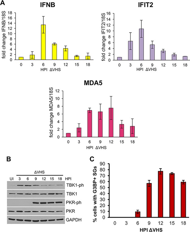
FIG 2.
Kinetics of induction of IFN-β and ISG mRNA, TBK1 and PKR activation, and SG assembly during ΔVHS infection. NHDFs were infected with ΔVHS at an MOI of 5 and collected at 3-h intervals. Uninfected (UI) cells were collected at 0 hpi. (A) mRNA levels of IFN-β and ISGs IFIT2 and MDA5 were analyzed by qRT-PCR from total RNA and normalized to 18S. The means ± the standard errors of the mean (SEM) of three independent experiments are plotted. (B) TBK1 (S172) and PKR (T446) phosphorylation were analyzed by immunoblotting of whole-cell lysates. (C) SG induction was quantified by counting the number of cells containing two or more G3BP1-stained SGs. More than 100 cells were counted for each time point, and the means ± the SEM of three independent experiments are plotted.

