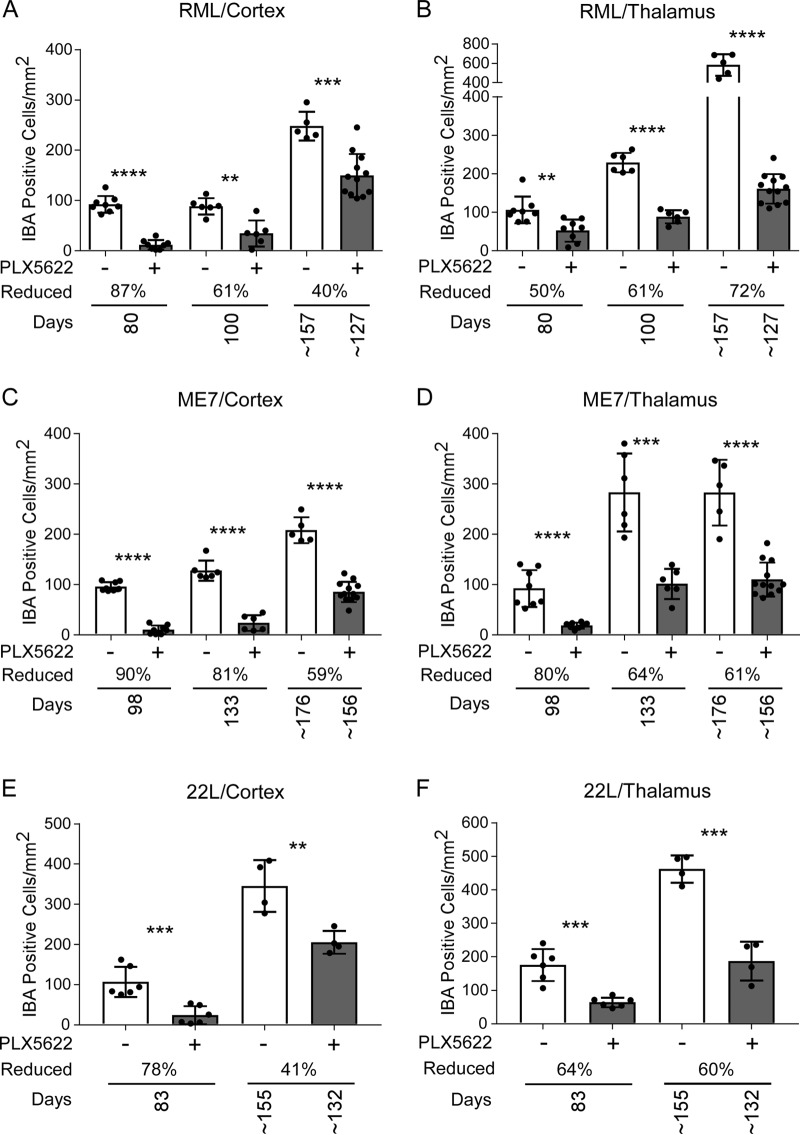
FIG 7.
Iba1-positive cells in cortex and thalamus during prion disease with PLX5622 treatment beginning at 14 dpi. Mice infected with scrapie strain RML, ME7, or 22L were fed control chow (−, white columns) or chow supplemented with PLX5622 (+, gray columns). Mice were euthanized at 80 dpi, at 100 dpi, and at clinical endpoint (∼157 dpi for control or ∼127 dpi for PLX5622) for RML-infected mice; at 98 dpi, at 133 dpi, and at clinical endpoint (∼176 dpi for control or ∼156 dpi for PLX5622) for ME7-infected mice; and at 83 dpi and at clinical endpoint (∼155 dpi for control or ∼132 dpi for PLX5622) for 22L-infected mice. See Table 1 for all clinical endpoint ranges. Similar paraffin-embedded sections of cortex (A, C, and E) and thalamus (B, D, and F) were fixed and stained with antibody to Iba1, and positive cells were enumerated and reported as the number of positive cells per square millimeter. The columns are the means, and each dot in the groups represents an individual mouse. The percent reduction with treatment for each time point relative to control is given. Error bars represent 1 standard deviation. Control and PLX5622-treated groups at each time were compared by unpaired t test: *, P ≤ 0.05; **, P ≤ 0.01; ***, P ≤ 0.001; ****, P ≤ 0.0001.

