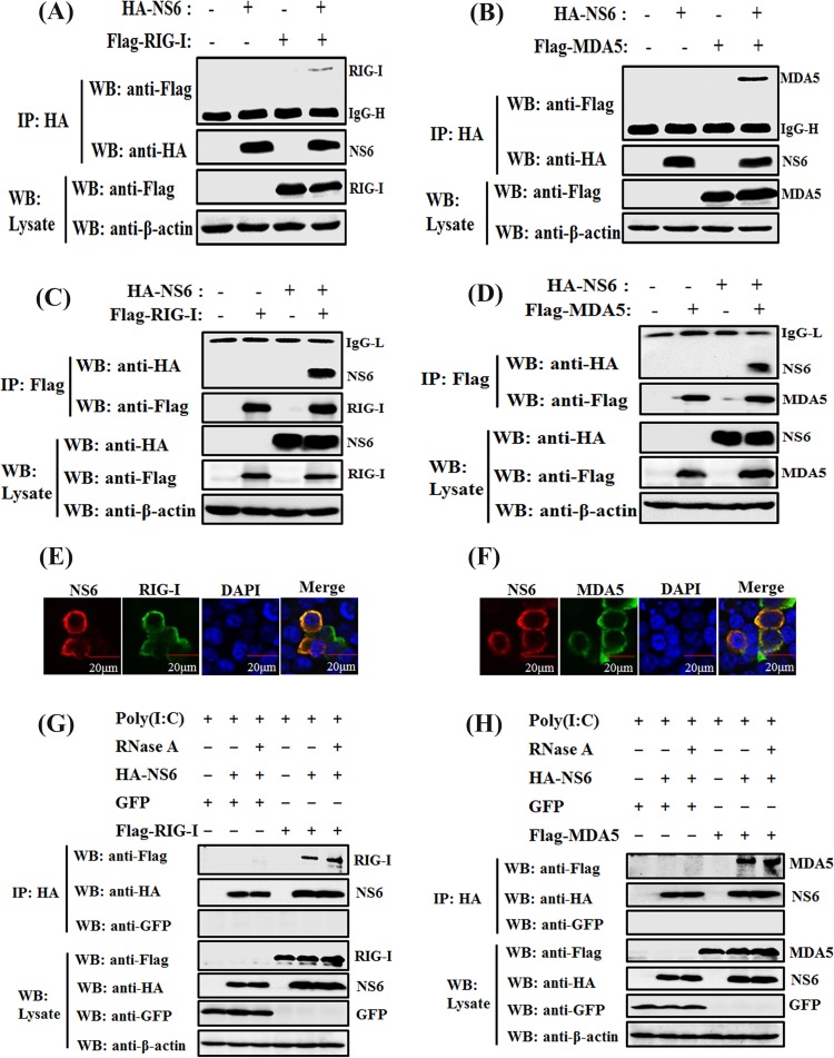
FIG 5.
NS6 interacts with both RIG-I and MDA5. (A to D) HEK-293T cells were cotransfected with pCAGGS-HA-NS6 and Flag-tagged RIG-I (A and C) or Flag-tagged MDA5 (B and D), respectively. At 28 h after transfection, cells were lysed and subjected to immunoprecipitation analysis with anti-HA (IP: HA) or anti-Flag (IP: Flag) antibody. The whole-cell lysates (WCL) and immunoprecipitation (IP) complexes were analyzed via Western blotting using anti-Flag, anti-HA, or anti-β-actin antibodies. (E and F) HEK-293T cells were cotransfected with pCAGGS-HA-NS6 and Flag-tagged RIG-I (E) or MDA5 (F). At 28 h after transfection, the cells were fixed for immunofluorescence assays to detect NS6 protein (red) and RIG-I or MDA5 (green) with anti-HA and anti-Flag antibodies, respectively. (G and H) HEK-293T cells were transfected with pCAGGS-HA-NS6 and pEGFP-C1, Flag-tagged RIG-I (G), or MDA5 (H) expression plasmids for 24 h, followed by the transfection of poly(I·C). Cells were lysed 36 h after transfection, and the clarified supernatants were left untreated or were treated with RNase A (50 μg/ml). The samples were then subjected to immunoprecipitation assays using anti-HA MAb (IP: HA). Cell lysates and immunoprecipitated complexes were subjected to Western blot assays as described for panels A and B.

