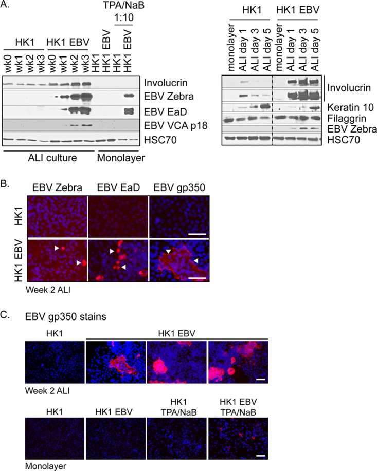
FIG 2 .

Induction of EBV lytic proteins in HK1-EBV ALI culture. (A) Immunoblot analysis for the expression of differentiation markers (involucrin, keratin 10, and processed filaggrin) and EBV lytic proteins (zebra, EaD, and VCA p18) in EBV-infected and uninfected ALI-cultured HK1 cell lines. Monolayer HK1 and HK1-EBV cultures with or without TPA/sodium butyrate induction were included for comparison and loaded at 1/10th of total protein lysates. Data for different antibodies are separated by horizontal white lines, and results from the same gel are grouped by a black border. Gels with intervening lanes that were cropped for labeling purposes are indicated by a vertical dotted line. (B) Immunofluorescence staining of HK1 and HK1-EBV cells at week 2 of ALI culture for the EBV lytic antigens zebra, EaD, and gp350 (red). (C) To reflect the frequency of reactivation, three representative fields of view are shown for the gp350 stain of HK1-EBV ALI cultures. For comparison, monolayer cultures were treated with TPA (200 nM) and sodium butyrate (5 mM) for 3 days to induce reactivation. Nuclei were counterstained with DAPI (blue). Scale bar, 50 µm. Images were acquired on an Olympus Provis epifluorescence microscope.
