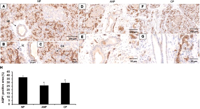
FIGURE 6.

Expression of AQP1 in patients with acute and chronic pancreatitis. Representative histological images show expression and localization of AQP1 in normal human pancreas (NP, A–C) acute necrotizing pancreatitis (ANP, D,E) and chronic pancreatitis (CP, F,G) samples. AQP1 immunoreactivity was detected in inter/intralobular and intercalated ducts, in the acinar and centroacinar cells of the normal pancreas. In contrast, expression of AQP1 strongly decreased in ANP and CP. id, interlobular duct; ic, intercalated duct; ca, centroacinar cells. (H) Intensity of diaminobenzidine staining was measured in pancreas samples by ImageJ software and expressed as percentage of total pancreatic area. ∗p ≤ 0.05 vs. normal pancreas, n = 10–12.
