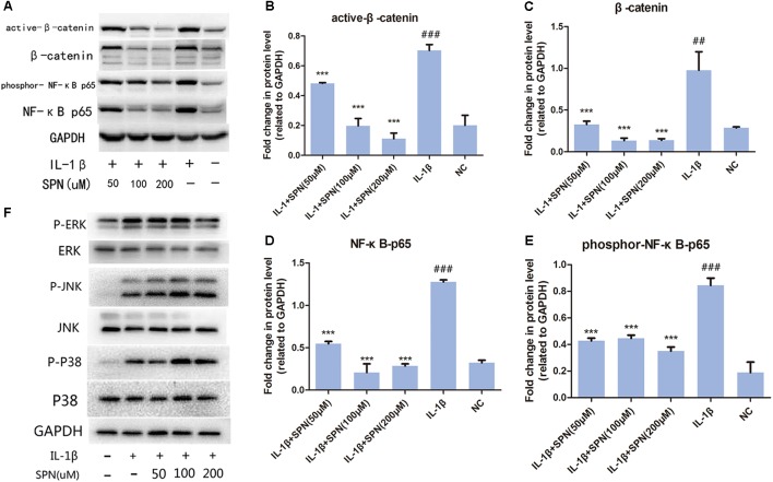
FIGURE 4.
Effect of SPN on IL-1β-induced activation of NF-κB and wnt/β-catenin signaling in rat chondrocytes. (A–E) Chondrocytes were treated with different concentrations of SPN for 6 h, with or without IL-1β (5 ng/ml). The levels of active-β-catenin, β-catenin, NF-κB p65, and GAPDH, as well as the phosphorylation level of NF-κB p65 were assessed by Western blotting. GAPDH was used as an endogenous control (n = 3 per group). (F) Chondrocytes were pretreated with different concentrations of SPN for 2 h, and then incubated with or without IL-1β (5 ng/ml) for 30 min. The levels of ERK, JNK, and P38, as well as their phosphorylation levels were assessed by Western blotting (n = 3 per group). Significance was calculated by one-way ANOVA with post hoc Tukey’s multiple comparisons test. ∗∗∗p < 0.001 versus the IL-1β+SPN (0 μM) group. ##p < 0.01, ###p < 0.001 versus the NC group.

