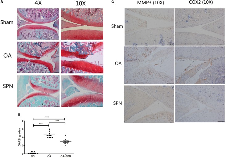
FIGURE 8.

Effect of SPN treatment on cartilage degeneration in a rat model of OA. ACL and MM-transected rats were injected intra-articularly with SPN (200 μM) every 7 days. (A) Rats were sacrificed to isolate the knees for analysis after 8 weeks of treatment. 4% paraformaldehyde-fixed knees were decalcified and embedded in paraffin, then sectioned at 5 μm thickness. Sections of the interior joint were stained with safranin O-fast green (SO). (B) The OARSI grading system (0–6) was used to evaluate the sections. (C) Inflammatory factors (MMP3 and COX2) were detected by immunohistochemistry (n ≥ 10 per group). Significance was calculated by one-way ANOVA with post hoc Tukey’s multiple comparisons test. ∗∗∗p < 0.001.
