
Figure 6.
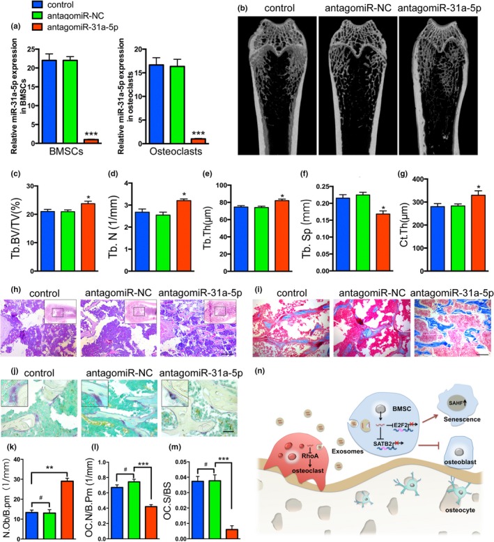
Injection of antagomiR‐31a‐5p into the bone marrow cavity stimulates bone formation and reduces osteoclastogenesis in aged rats. (a) qRT‐PCR analysis of levels of miR‐31a‐5p expression in BMSCs and osteoclasts of rats with antagomiR‐31a‐5p. (b) Micro‐CT images and quantitative CT analysis (c–g) were performed in the distal femur from 15‐month‐old rats treated with control, antagomiR‐NC and antagomiR‐31a‐5p. (h and i) Representative images of HE and Masson's trichrome staining exhibited that bone structure and (k) osteoblasts were significantly increased in aged rats injected with antagomiR‐31a‐5p. (j) Trap staining of distal femur in the three groups and (l) quantification of OC.N/B.Pm (osteoclast number per bone perimeter) and (m) OC.N/BS (osteoclast number per bone surface) indicated that osteoclast numbers were significantly decreased on the bone surface with antagomiR‐31a‐5p treatment compared to those with antagomiR‐NC and control treatment. (n) A schematic working model for miR‐31a‐5p function in the regulation of BMSCs and osteoclast. # p > 0.05, *p < 0.05, **p < 0.01, ***p < 0.001. Scale bars: 200 μm (h); 100 μm (i); 50 μm (j). Data are presented as the mean ± SD, n = 3