Figure 1.

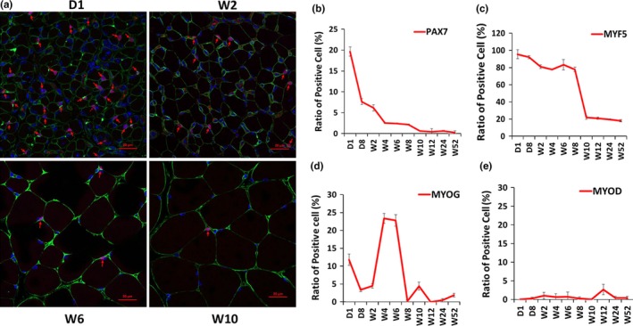
Expression patterns of myogenic factors in skeletal muscle development. Paraffin section immunofluorescence was performed to test the expression patterns of PAX7, MYF5, myogenin, and MYOD in the gastrocnemius muscle of mice at different developmental stages. (a) Confocal images of the immune stain of PAX7 (red) and laminin (green) proteins. D1, W2, W6, and W10 are shown as representatives. Nucleus was stained with DAPI (blue). PAX7‐positive cells are marked with red arrows. Scale bars: 20 μm. Magnification: 400×. (b) Change in the ratio of PAX7+ cells at 10 time points. (c) Change in the ratio of MYF5+ cells at 10 time points. (d) Change in the ratio of myogenin+ (MYOG) cells at 10 time points. (e) Change in the ratio of MYOD+ cells at 10 time points. The number of positive cells is presented as mean ± SEM (12 random fields are captured for each treatment group)
