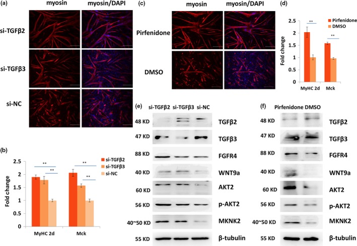
Figure 5.

TGFβ2 and TGFβ3 negatively regulate the differentiation of satellite cells. (a) Immunofluorescence staining of myosin (red) in the 24‐hr differentiated satellite cells when Tgfβ2 or Tgfβ3 was inhibited using RNAi. Nucleus was stained with DAPI (blue) Scale bars: 200 μm. Magnification: 100×. (b) qPCR results of the expression change in MyHc2d and Mck when TGFβ2 or TGFβ3 was inhibited using siRNA. (c) Immunofluorescence staining of myosin (red) when TGFβ2 and TGFβ3 were inhibited using pirfenidone. Nucleus was stained with DAPI (blue) Scale bars: 200 μm. Magnification: 100×. (d) qPCR results of the expression change in MyHC2d and Mck when TGFβ2 and TGFβ3 were inhibited using pirfenidone. (e, f) Western blot results of TGFβ2, TGFβ3, WNT9a, FGFR4, AKT2, p‐AKT2, and MKNK2 in proliferative satellite cells when TGFβ2 and TGFβ3 was inhibited using siRNA (e) or pirfenidone (f). Tubulin was used as the internal control for qPCR and western blot. Triplicate samples were analyzed for each treatment, and the results are presented as the mean ± SEM *p < 0.05; **p < 0.01
