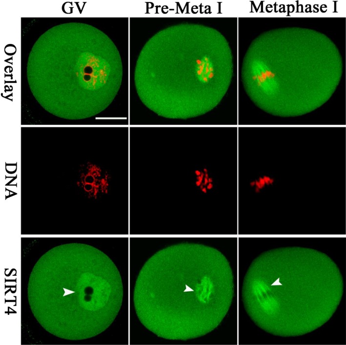
Figure 1.

Cellular localization of SIRT4 in mouse oocytes. Oocytes at GV, Premetaphase I, and metaphase I stages were immunolabeled with SIRT4 antibody (green) and counterstained with PI to visualize DNA (red). Arrowheads point to the accumulated SIRT4 signals. Scale bar: 25 μm
