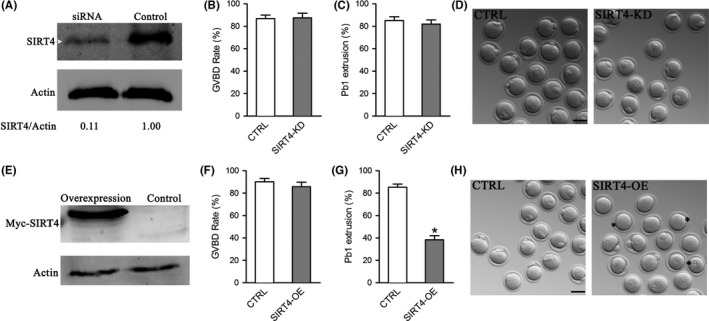
Figure 2.

Effects of SIRT4 expression on the meiotic progression of mouse oocytes. Fully grown oocytes injected with SIRT4‐cRNAs or SIRT4‐siRNAs were arrested in medium with milrinone for 20 hr, washed in milrinone‐free medium, and then cultured in vitro for the following analysis. (A) Efficiency of SIRT4 knockdown (SIRT4‐KD) after siRNA injection was verified by immunoblotting. Actin served as a loading control. Band intensity was calculated using ImageJ software, and the ratio of SIRT4/Actin expression was normalized and values are indicated. (B–C) Quantitative analysis of GVBD and Pb1 extrusion rate in control (n = 137) and SIRT4‐KD (n = 112) oocytes. (D) Representative images of control and SIRT4‐KD oocytes. (E) Efficiency of SIRT4 overexpression (SIRT4‐OE) after cRNA injection was verified by immunoblotting. (F–G) Quantitative analysis of GVBD and Pb1 extrusion rate in control (n = 120) and SIRT4‐OE (n = 122) oocytes. (H) Representative images of control and SIRT4‐OE oocytes. Asterisks indicate the oocytes that fail to extrude polar body. The graph shows the mean ± SD of the results obtained in three independent experiments. *p < 0.05 vs. controls. Scale bar: 100 μm
