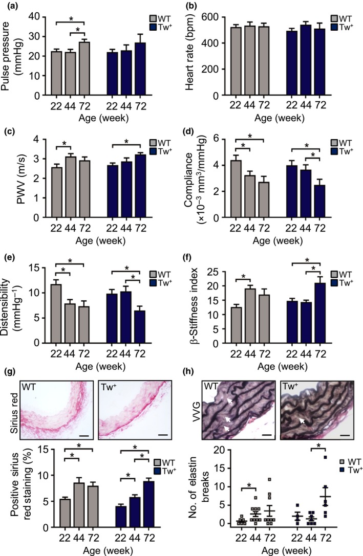
Figure 5.

Tw+ mice have delayed vascular aging. (a–f) Pulse pressure, heart rate, aortic pulse wave velocity (PWV), and carotid artery compliance, carotid artery distensibility and carotid β‐stiffness index in WT and Tw+ mice aged 22–72 wk (n = 6–9), (g) representative images of aorta from 44‐wk‐old WT and Tw+ mice stained with Sirius Red and quantification of the percentage positive staining (scale bar = 50 μm) and (h) representative images of aorta from 44‐wk‐old WT and Tw+ mice stained with Verhoeff–van Gieson (VVG) showing elastin breaks (white arrows) and quantification of the number of elastin breaks. Scale bar = 20 μm (n = 3–7). WT (grey labels); Tw+ (blue labels). Data are means ± SEM. *p < .05 using ANOVA with Tukey post‐test
