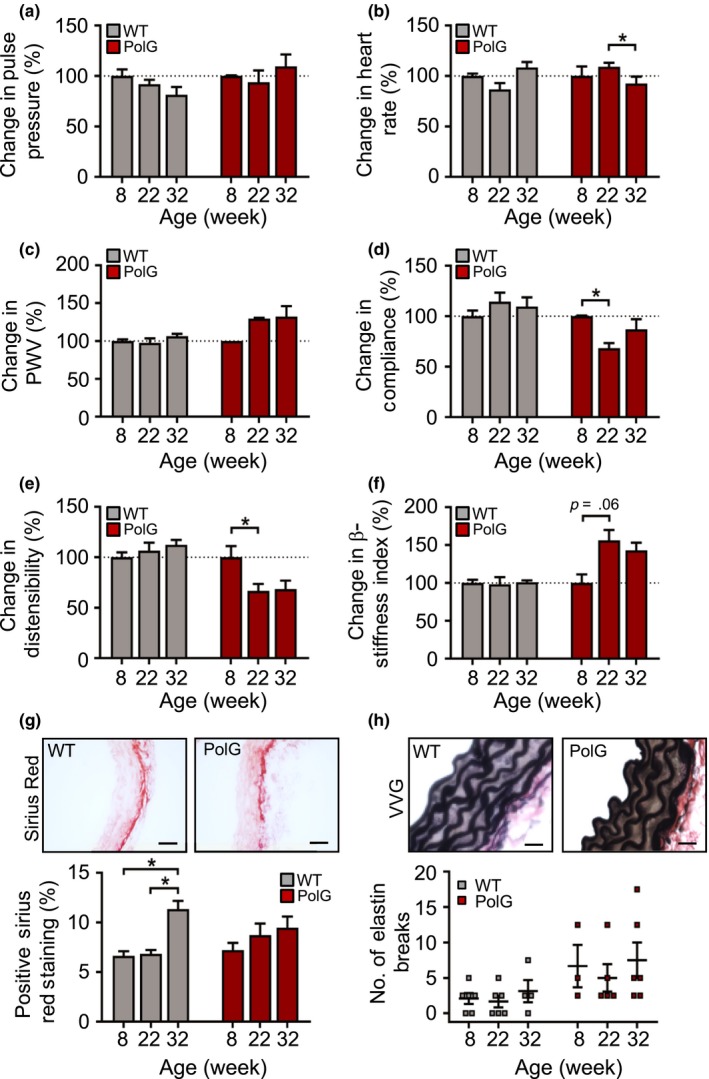
Figure 6.

PolG mice have accelerated vascular aging. (a–f) Pulse pressure, heart rate, aortic pulse wave velocity (PWV) and carotid artery compliance, carotid distensibility and carotid β‐stiffness index in WT and PolG mice aged 8–32 wk relative to 8‐wk‐old mice (n = 5–9), (g) representative images of aortas from 22‐wk‐old WT and PolG mice stained with Sirius Red and quantification of positive staining (scale bar = 50 μm) and (h) representative images of aortas from 8‐wk‐old WT and PolG mice stained with Verhoeff–van Gieson (VVG) for measuring elastin breaks (scale bar = 20 μm) (n = 4–6). WT (grey labels); PolG (red labels). Data are means ± SEM. *p < .05 using ANOVA with Tukey post‐test
