Figure 5.

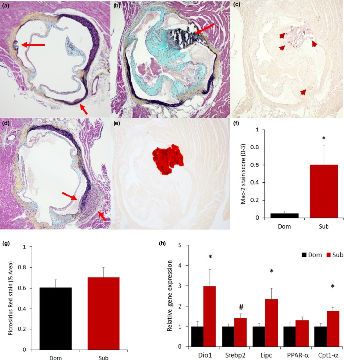
Early‐stage atherosclerotic lesions of the aortic sinuses are found exclusively in subordinate mice. Exemplar slides stained with Movat's staining showing the presence of rupture (a, top red arrow), immune cells infiltration (a, bottom red arrow, b and d) as well as calcification as confirmed by Alizarin stain (b, e). Inflammation was verified by Mac‐2 staining (c, red arrow) and quantified with a Mac‐2 score [f, F(1,14) = 5.76, p < .05]. (g) Quantification of fibrosis in the mouse myocardium using Picrosirius Red stain (representative images are shown in Figure S6). (h) Liver expression of lipid cholesterol metabolism markers [iodothyronine deiodinase 1 (Dio1), sterol regulatory element‐binding protein 2 (Srebp2), lipase C (Lipc), peroxisome proliferator‐activated receptor alpha (PPAR‐α), carnitine palmitoyltransferase 1α [(Cpt1α)]. N = 8/group *p < .05, # p < .07
