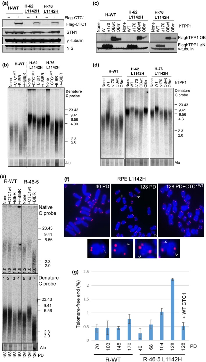
Figure 3.

CTC1 L1142H promotes telomere elongation by telomerase. (a) Determination of the expression of WT Flag‐CTC1 and endogenous STN1 in the indicated cell lines by western analysis. (b) TRF Southern analysis of telomere length in WT or mutant HCT116 cells reconstituted with either WT CTC1 or cultured in the presence of 10 μM of the telomerase inhibitor BIBR. Cells were first passaged for 120 PD, reconstituted with WT CTC1 or treated with BIBR, and maintained for another 120 PD. +/−BIBR: cells were maintained in the presence of 10 μM BIBR for 60 PD, then maintained for another 60 PD after discontinuing BIBR treatment. (C) Expression levels of WT TPP1, TPP1∆170 or TPP1‐OBWT or OBRR (K166R; K167R mutations) in HCT116 cells by western analysis. (d) Analysis of total telomeric DNA in WT or mutant HCT116 cells expressing either WT TPP1, TPP1∆170 or TPP1‐OBWT or OBRR. Cells expressed TPP1 constructs for 60 days before undergoing telomere length analysis by TRF Southern. Alu was used as a DNA loading control. (e) Telomere length analysis of single‐stranded (ss) (top panel) and total telomere length (bottom panel) in cells of the indicated genotypes. PD: population doublings. +CTC1: WT CTC1 was expressed for 120 days before the cells were harvested for telomere length analysis. +BIBR: cells were treated with 10 μM of the telomerase inhibitor BIBR. Numbers in native gel refer to ratio of overhang signal intensity to total telomere intensity. (f) Metaphase spreads revealing sister telomere loss in RPEL 1142H cells at the indicated PD. White arrowheads point to STLs. In one experiment, WT CTC1 was expressed in RPEL 1142H cells at PD 54 and then passaged for an additional 74 PD. (g) Quantification of the percentage of sister telomere loss in WT RPE or RPEL 1142H cells with the indicated PD when harvested
