
Figure 3.
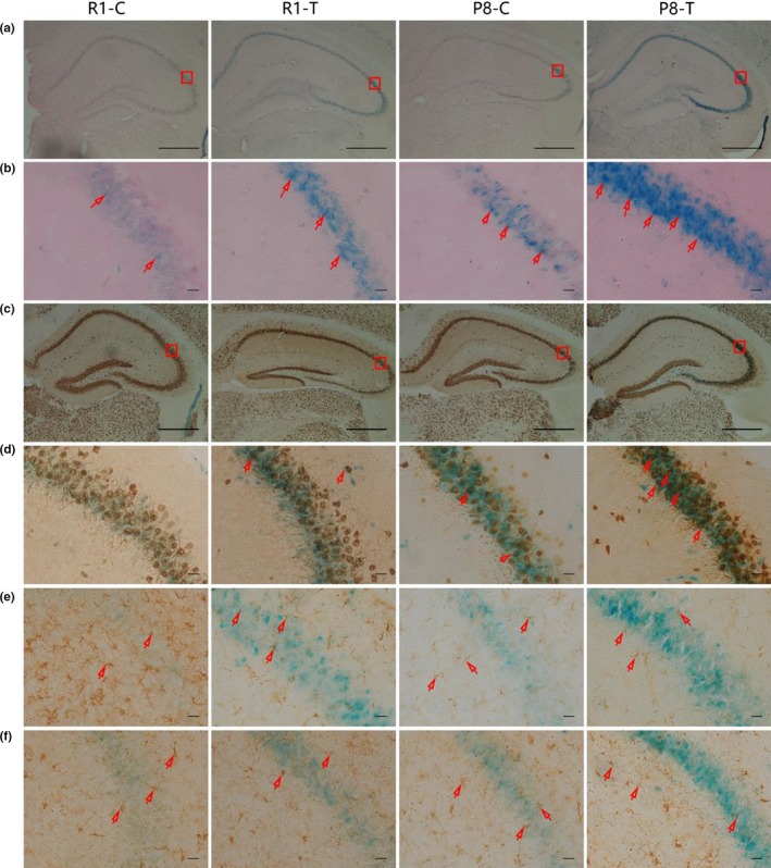
Trimethylamine‐N‐oxide (TMAO) induced the aging process in the hippocampal CA3 region. (a and b) SA‐beta‐GAL was detected in the hippocampal CA3 region. The blue cells indicated SA‐beta‐GAL‐positive cells. (c–f) Double staining with SA‐beta‐GAL and immunohistochemical staining for senescent cells. (c and d) Cells positive for SA‐beta‐GAL and NeuN (a marker of neurons, the red arrow indicates senescent neurons) were found in the CA3 region of the hippocampus. (e) Cells negative for SA‐beta‐GAL but positive for GFAP (a marker of astrocytes, the red arrow indicates astrocytes) were found in the CA3 region of the hippocampus. (f) Cells negative for SA‐beta‐GAL but positive for Iba1/AIF1 (a marker of microglia, the red arrow indicates microglia) were found in the CA3 region of the hippocampus. These results suggested that TMAO could promote cell senescence in the hippocampal CA3 region, and the senescent cells were primarily neurons (n = 6 each group, scale bar: b, d, e, f = 100 μm, a, c = 50 μm)