
Figure 6.
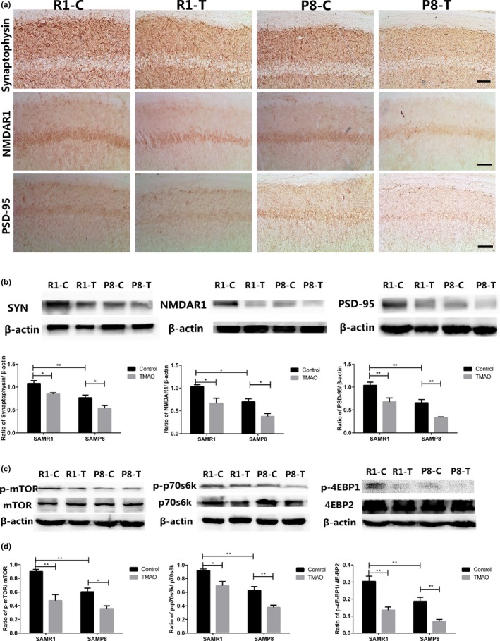
Trimethylamine‐N‐oxide (TMAO) reduced the expression of synaptic plasticity‐related proteins by down‐regulating the activity of the mTOR signalling pathway. (a) Synaptophysin (SYN), postsynaptic density 95 (PSD‐95) and N‐methyl‐d‐aspartate 1 (NMDAR1) were shown by immunohistochemical staining in the hippocampus. The expression levels of SYN, NMDAR1 and PSD‐95 were significantly decreased in the P8‐C group compared with the R1‐C group. There was a significant decrease in the expression of these synaptic‐associated proteins in the TMAO groups compared with their control groups (scale bar = 50 μm). (b) The expression levels of SYN, NMDAR1 and PSD‐95 were assayed by Western blotting. To examine the possible mechanism involved in the decline of synaptic‐associated proteins, three proteins (mTOR, p70s6k and 4EBP2) of the mTOR signalling pathway were assessed by RT–PCR and Western blotting. (Figure S3A) The mRNA levels of mTOR, p70s6k and 4EBP2 were not significantly different in the groups assessed by RT–PCR. (c) Phosphor‐mTOR/mTOR, phosphor‐p70s6k/p70s6k and phosphor‐4EBP1/4EBP2 were monitored by Western blotting. β‐Actin was used as a loading control. The bands in the Western blotting were scanned, and the ratios of optical density of specific bands and β‐actin are illustrated. TMAO inhibited the active form of mTOR, p70s6k and 4EBP2 (phosphorylated). Data are shown as the mean ± SEM (n = 6 each group, two‐way ANOVA, comparisons between two groups were made using Tukey's multiple comparison test. **p < .01). The samples derived from the same experiment and the blots were processed in parallel