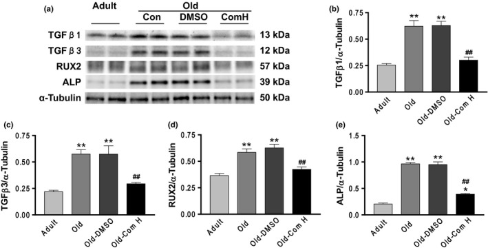
Figure 6.

Aging increased arterial TGF‐β1, TGF‐β3, RUNX2, and ALP expressions which can be abolished by compound H. (a) Western blots analysis of TGF‐β1, TGF‐β3, RUNX2, and ALP. (b) Quantification of TGF‐β1 protein levels. (c) Quantification of TGF‐β3 protein levels. (d) Quantification of RUX2 protein levels. (e) Quantification of ALP protein levels. Data are expressed as mean ± SE and analyzed by one‐way ANOVA. n = 4. *p < .05, **p < .01 vs. adult mice; ## p < .01 vs. old mice
