
Figure 3.
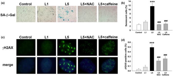
Prosenescent effect of L5 on human aortic endothelial cells (HAECs) in vitro. HAECs were treated with phosphate‐buffered saline (PBS) (control), L1 (30 μg/ml), L5 (30 μg/ml), L5 + NAC (5 mM), or L5 + caffeine (1 mM) for 72 hr (n = 4 independent experiments per treatment group). (a) SA‐β‐Gal staining in HAECs from each treatment group. Positively stained cells were quantified and are shown as percentages of the total number of cells in (b). (c) Immunofluorescence staining showing γH2AX foci in HAECs from each treatment group. Positively stained cells were quantified and are shown as percentages of the total number of cells in (d). DAPI (blue) counterstaining shows the nuclear localization of γH2AX. ***p < 0.01 vs. control; ### p < 0.01 vs. L5