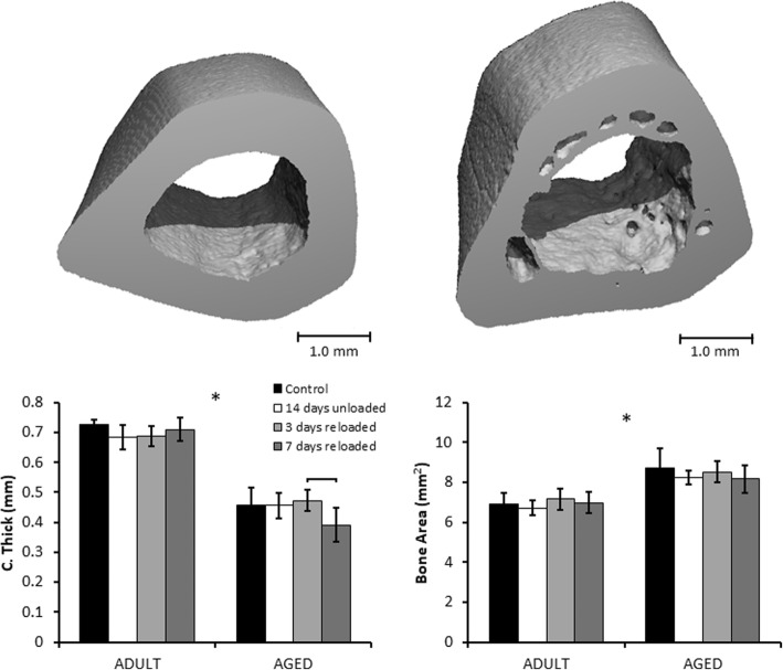
Fig. 4.
Mid Femoral Diaphysis μCT Results. Representative samples of regions of interest in Adult (left) and Aged (right) femurs. Aged rats had lower average cortical thickness than Adult rats, but greater bone area and total cross-sectional area. Neither age group exhibited changes in cortical structure due to hindlimb unloading or reloading. Brackets indicate significant differences between groups (p < 0.05). Asterisks (*) indicate a main effect of age in a parameter between Adult and Aged animals (p < 0.05)

