
Figure 1.
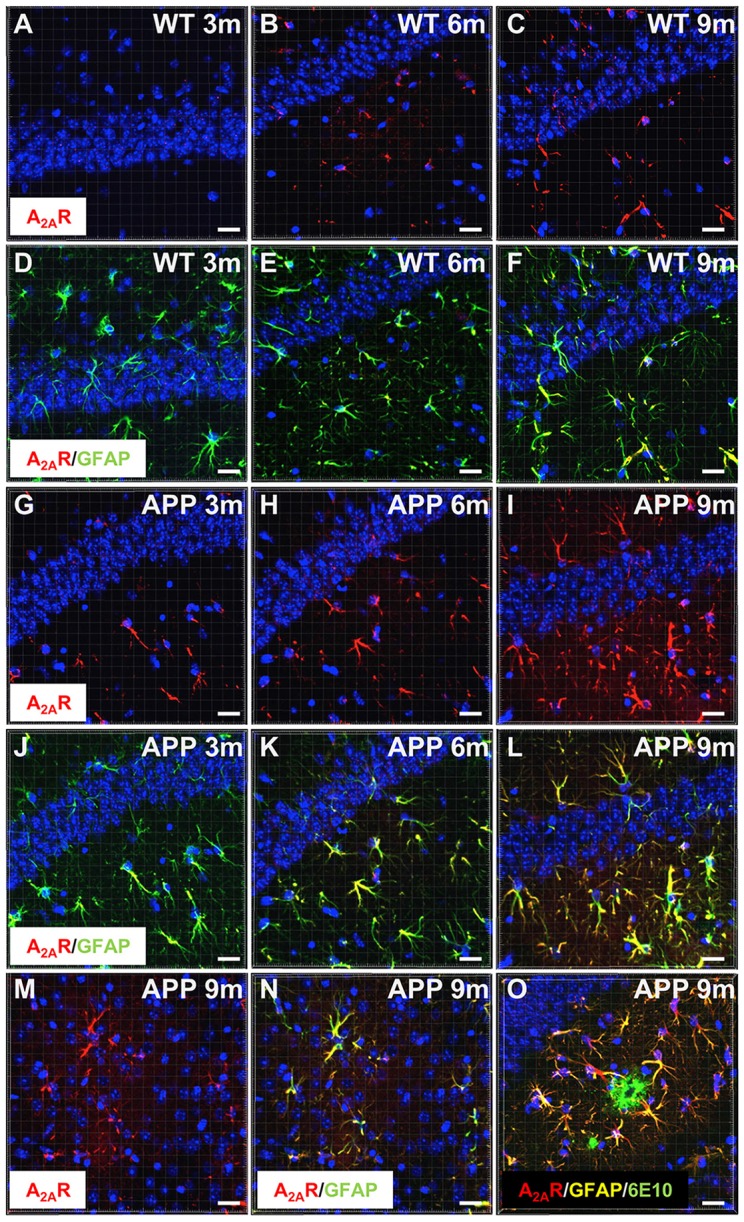
Astrocytic upsurge of A2ARs in APP/PS1 mice. Representative photomicrographs of hippocampal immunostaining for the A2A receptor (A2AR; red) (A–C,G–I) and merged with the astrocyte marker GFAP (green) (D–F,J–L) from WT mice (A–F) and the APP/PS1 mice (G–L), at different ages: 3 months (A,D,G,J), 6 months (B,E,H,K), and 9–10 months (C,F,I,L). Representative photomicrographs of A2AR expression (red) (M) and the merged with GFAP marker (green; N) in the cortex of 9-month-old APP/PS1 mice. Representative photomicrograph of A2AR expression (red) GFAP (yellow) and 6E10-positive amyloid plaque marker (green) in the hippocampus of 9 month-old APP/PS1 mice (O). Cell nuclei were labeled with DAPI (blue). Scale bar = 20 μm.