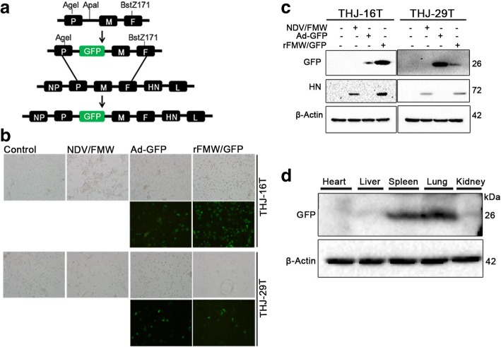
Fig. 1.
Construction and identification of the recombinant rFMW/GFP. a PCR amplification of the DNA construct using ApaI-tagged primers was carried out and subsequently introduced into the Anti-sense cDNA of the NDV strain, FMW. b THJ-16 T and THJ-29 T cells were infected with vehicle, NDV/FMW (10 MOI), adenovirus-GFP-Tag or rFMW/GFP (10 MOI) and imaged by fluorescence microscopy. Live cell imaging using bright-field and fluorescence microscopy was recorded at 24 h post-infection. c Protein levels of GFP-tagged and HN proteins were analyzed by immunoblotting (IB). d rFMW/GFP (1 × 10⁷ TCID50 per dose) was i.v. injected into BALB/c mice. Mice were sacrificed 24 h post virus injection. Heart, liver, spleen, lungs and kidneys were harvested and GFP protein expression was assessed by IB. β-actin was used as a control for equal loading. All IB experiments were performed twice

