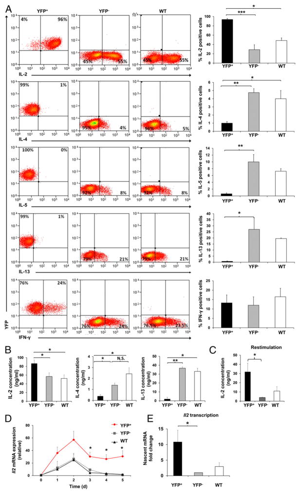
FIGURE 2. Increases in IL-2 and decreases in Th2 cytokine expression in HuR-ablated CD4+ T cells.
(A) YFP+ (HuR-KO), YFP− (endogenous control), or WT (HuRfl/fl control with no Cre) CD4+ T cells were stimulated under nonpolarizing conditions with plate-bound anti-CD3 and anti-CD28. On day 5 of activation, cells were harvested, stimulated with PMA and ionomycin for 5 h, and stained for intracellular cytokines. (B) IL-2, IL-4, and IL-13 levels in the culture supernatant under nonpolarizing conditions on day 5 postactivation of YFP+, YFP−, and WT CD4+ T cells, as measured by ELISA. (C) IL-2 levels in activated YFP+, YFP−, and WT CD4+ T cells after restimulation with PMA and ionomycin for 5 h, as detected by ELISA. (D) RT-PCR analysis of Il2 mRNA in YFP+, YFP−, and WT CD4+ T cells activated under

