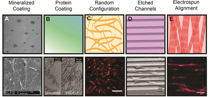
Figure 3. Biomaterial topography instructs MSC adhesion and alignment to guide cell phenotype.

(A) Top: Polymer coating with biomineral increases protein adsorption, cell adhesion, and spreading. Bottom: MSC spreading on biomineralized PLG films.10 (B) Top: Coating with functional proteins regulates integrin engagement. Bottom: MSC adhesion to RGD-linked elastin-like peptide-coated titanium.137 (C) Top: Randomly configured nanofibers in ECM for increased surface area of cell adhesion. Bottom: MSCs on decellularized MSC-secreted ECM undergo osteogenic differentiation.2 (D) Top: Defined surface structures guide cell spreading and orientation. Bottom: MSCs on micropatterned polyimide undergo osteogenic differentiation.138 (E) Top: Aligned electrospun nanofibers guide cell orientation and enhance FAK signaling. Bottom: MSCs on aligned PLLA/PCL exhibit alignment at Day 1 and subsequent increases in myogenic differentiation.64 Reproduced with permission from Refs. 2, 10, 64, 137-138 Copyrights 2010 John Wiley and Sons, 2016 Elsevier, 2016 American Chemical Society, 2015 Elsevier, 2013 John Wiley and Sons.
